Calcic Clinopyroxene


Occurrence
Augite, the most common clinopyroxene, is found in mafic to intermediate igneous rocks and their metamorphic equivalents. Diopside, a Ca-Mg clinopyroxene is found mostly in marbles.

Composition
Clinopyroxenes have general formula (Ca,Mg,Fe,Na)(Mg,Fe,Al)(Si,Al)2O6. Most clinopyroxenes in igneous and metamorphic rocks (generally termed augite) can be thought of as (Ca,Mg,Fe)2Si2O6, with small amounts of Al, Mn and Na substituting for other elements. The most significant clinopyroxene end member is diopside, CaMgSi2O6. Its Fe equivalent, CaFeSi2O6 is termed hedenbergite. Near end-member diopside is found in some marbles. Diopside and hedenbergite are the most dominant components in most augite.

Identification
The keys to identifying calcic clinopyroxene are normally its high relief, pale green (sometimes clear or light brown) color, middle second-order interference colors, and near 90o cleavage seen in some views. Distinguishing the different pyroxenes and olivine can sometimes be difficult.

Important properties:
 ·Color - Common augite is generally light green; Fe-free clinopyroxene such as diopside may be clear; other varieties may be light brown or yellow. Darker color and weak pleochroism are characteristic of more Fe-rich varieties.
 ·Relief is moderate to faily high; clinopyroxene typically stands out above feldspar, with which it is often associated.
 ·Interference colors - Interference colors range up to high second order.
 ·Appearance and cleavage -Well developed end sections may appear as blocky four- or eight-sided crystals; more typically forms stubby prisms or football shaped grains, or anhedral crystals and masses. Basal sections show cleavage angle near 90o. Longitudinal sections show one cleavage.
 ·Twinning and exsolution - Although many clinopyroxenes are neither twinned nor show exsolution, both features are common. Exsolved orthopyroxene may form fine lamellae in clinopyroxene; simple or polysynthetic twins may give a striped or herringbone appearance.
 ·Interference figure - 2V and optical sign vary with composition.

Similar minerals:
 ·Orthopyroxene has parallel extinction and lower birefringence.
 ·Pigeonite, a low-Ca high temperature clinopyroxene has lower 2V.
 ·Sodic pyroxenes are generally more strongly colored and have larger 2V.
 ·Amphiboles may appear similar but have characteristic near 60o-120o cleavage angles.
 ·Olivine lacks cleavage and has higher birefringence.


Diopside in a Dolmitic Marble

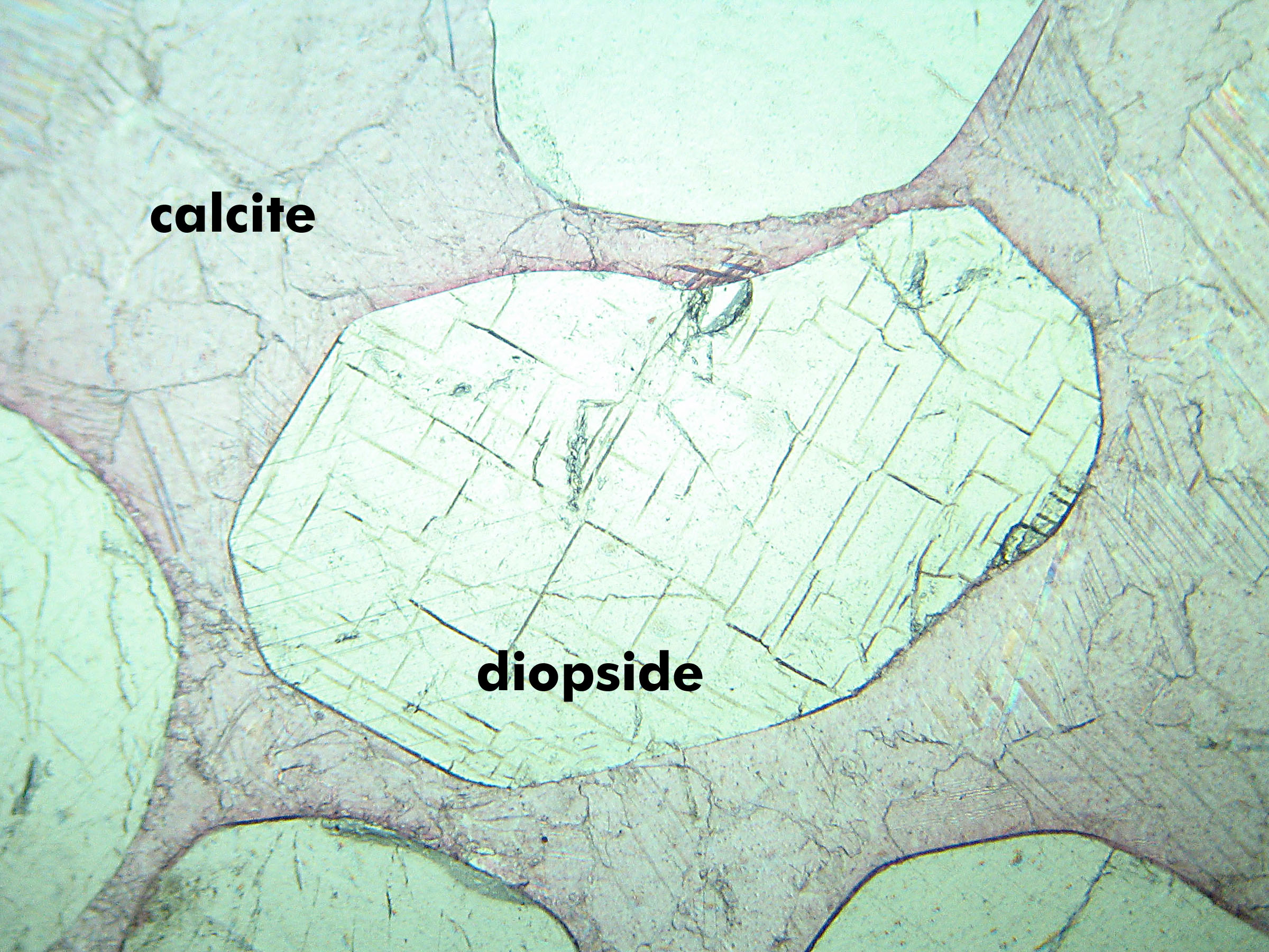
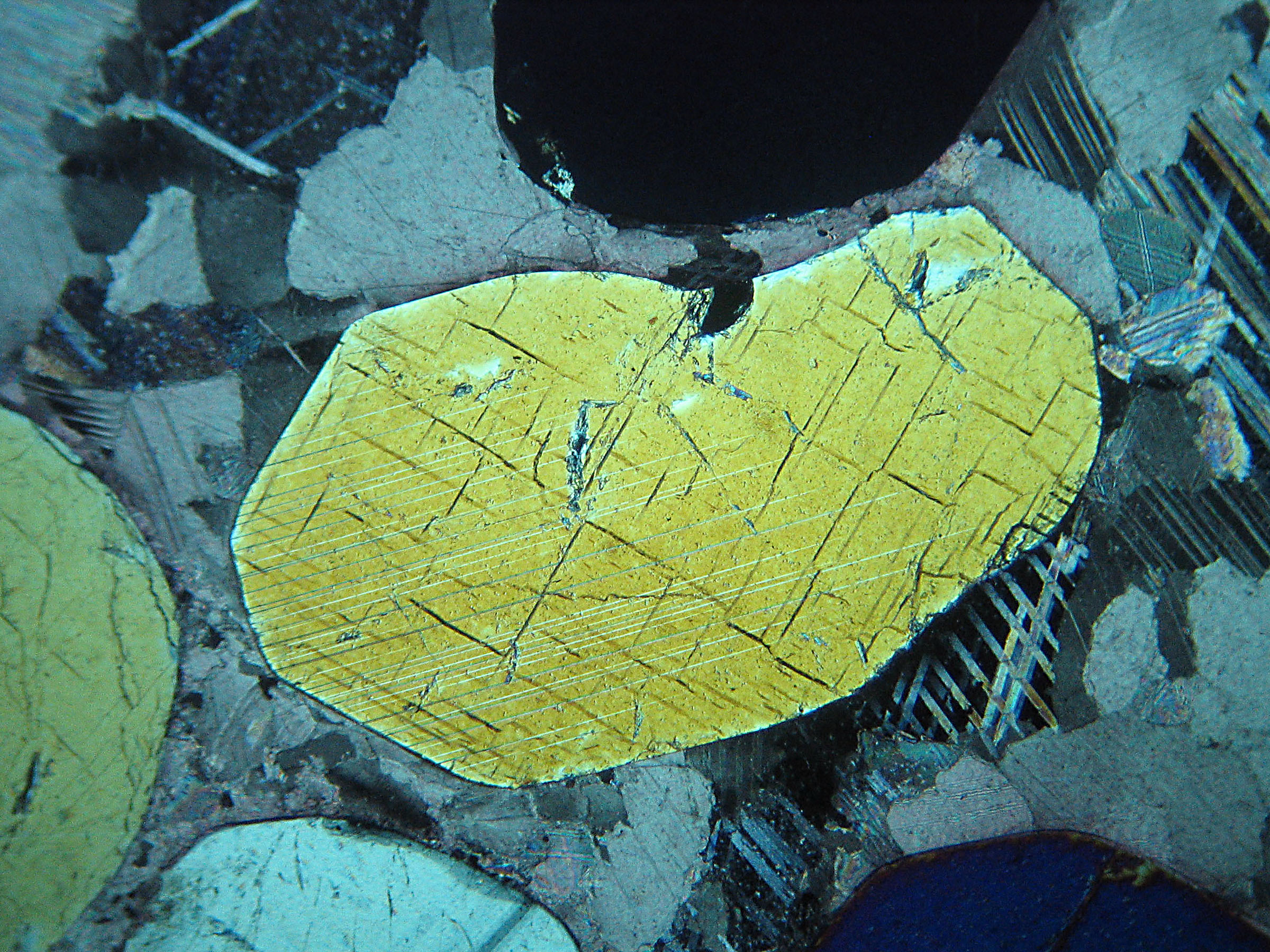
These photos show diopside in a marble from the Adirondack Mountains, New York. Field of view is about 2 mm. The football shaped diopside grain in the center shows classic near 90o angle between cleavages - diagnostic of pyroxene. It also shows incipient twinning (XP). Diopside's inteference colors range up to mid second order but in views that show two cleavages tend to be lower. This diopside is in a matrix of dolomite. Here the dolomite appears slightly pinkish because the thin section was etched and stained with alizarin red stain to help distinguish calcite from dolomite (calcite stains a darker red color). The dolomite is twinned and shows very high order interference colors; they appear as pastels, in places almost pearly white.


Clinopyroxene in a Spinel Peridotite Xenolith from Kilbourne Hole, New Mexico

These photos show mostly clinopyroxene (augite). A large grain of orthopyroxene, with slightly greater relief than the clinopyroxene, is on the left edge of the photos, and several grains of green (PP) isotropic (XP) spinel can be seen in the upper right.

The field of view is 4.5 mm.


Clinopyroxene in a Lherzolite from the Cima Volcanic Field, California

The photos (3 mm across) show mostly clinopyroxene. The large kite-shaped grain in the center is clinopyroxene. The high relief shrimp-shaped grain up and to the right of it (showing second order purply-red-orange interference colors), and the triangular grain with yellow-green interference colors below it, are olivine. Several fractured grains showing first order white to gray interference colors are orthopyroxene. One small flake of brown biotite (PP) can be seen in the lower center.


Microcline and Hedenbergite in a Syenite from Quebec

The photos show hedenbergite (Fe-rich calcic clinopyroxene) with microcline. The pyroxene has higher relief than the microcline and a slight greenish color (PP). It also has poorly developed cleavage. In the XP view, the pyroxene shows 2nd order interference colors; the microcline only first order grays and white. The microcline also displays well developed twins.

The field of view is about 2.5 mm across.


Olivine, Augite, and Plagioclase in a Porphyry from Near Fish Lake, Oregon

This view shows three kinds of phenocrysts in a groundmass of mostly plagioclase, magnetite (opaque) and clinopyroxene. The phenocrysts are lathes of plagioclase, several more equant grains of olivine, and two grains of augite. In PP light all three are nearly clear, but the augite (hard to pick out in PP) has a slightly greenish tint.

In XP light, the olivine can be seen to have 2nd order pastel interference colors that vary within individual grains. The largest olivine grain, a quarter of the way down from the top center of the photo) shows red, violet, blue and green interference colors. The two largest augite grains are next to it - one up and to the right and one below and slightly left. Both augite grains show somewhat anomalous yelllow-brown interference colors.

The field of view is about 2.5 mm across.


Augite and Plagioclase in a Basalt from India

This view shows high relief light green augite (clinopyroxene), opaque magnetite, and clear plagioclase (PP). In the XP view the augite has up to mid second order interference colors and the plagioclase shows twinning in first order black-gray-white interference colors. The augite shows cleavage, and the angle between cleavages appears to be close to 90o in some grains.

The field of view is about 1.5 mm across.


  OmniUpdate